
Multimodal Imaging Platform Optimized for the Posterior Segment
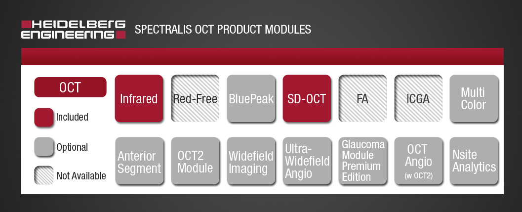
The SPECTRALIS® is an ophthalmic imaging platform with an upgradable, modular design. This platform allows clinicians to configure each SPECTRALIS to the specific diagnostic workflow in the practice or clinic.
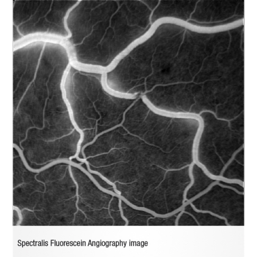
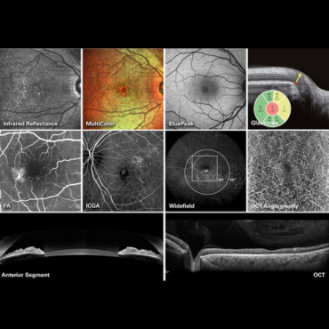
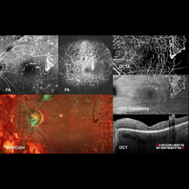
Multimodal imaging options include: OCT, multiple scanning laser fundus imaging modalities, widefield and ultra-widefield, scanning laser angiography and OCT angiography.
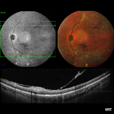
The SPECTRALIS® OCT imaging platform combines scanning laser fundus imaging with simultaneously acquired OCT.
As an expandable platform, it can be upgraded with additional scanning laser fundus imaging modalities, such as BluePeak autofluorescence and MultiColor, as well as advanced modules such as OCT2 and the Glaucoma Module Premium Edition.
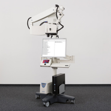
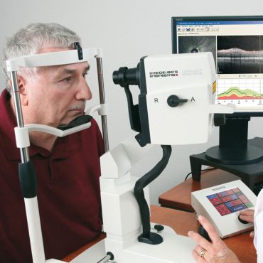
The SPECTRALIS OCT is also available with an optional panning camera head and an external touch panel control for easy adjustment and acquisition.
FEATURES:
Upgradeable Platform
SPECTRALIS® is a truly flexible and upgradeable imaging platform. Customize your SPECTRALIS precisely to your individual needs, confident in the knowledge that your OCT will grow with your clinic. As new technology becomes available, simply add new imaging modalities to your SPECTRALIS, providing you with additional information to enhance clinical decision-making and preserving patient data for precise follow-up.
Confocal Scanning Laser Ophthalmoscopy
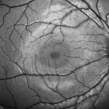
The confocal scanning laser ophthalmoscope (cSLO) in the SPECTRALIS® platform is an innovative technology for examining and imaging the retina and other eye structures.
Combining the selectivity of laser light with the pinpoint resolution of confocal scanning, the cSLO provides image detail and clarity not available from fundus photography.
The cSLO technology not only offers documentation of clinical findings but also often highlights critical diagnostic details not visible on traditional clinical ophthalmoscopy. Since cSLO imaging minimizes the effects of light scatter, it can be used effectively even in patients with cataracts.
Multimodality Diagnostics
The unique combination of imaging modalities provides the additional information you need to make confident clinical decisions. Use different, established and novel imaging techniques simultaneously to improve your understanding of different pathologies.
TruTrack Active Eye Tracking
TruTrack Active Eye Tracking is a patented imaging technology that utilizes two beams of light simultaneously to track and image the eye. Actively tracking the eye in real-time throughout image capture, mitigates the effects of eye motion, resulting in accurate OCT scan data.
Additional clinical benefits of TruTrack Active Eye Tracking are precise, automated retinal follow-up scanning, retinal thickness measurement reproducibility to 1 micron, and excellent image quality throughout the volume scan. This superior performance allows a more effective management of patients, even in challenging cases with very small changes over time.
AutoRescan
Using the SPECTRALIS® fundus image like a map, the AutoRescan function automatically places follow-up scans in precisely the same position visit after visit. Accurate, automatic placement of follow-up scans is important for optimizing patient flow and for confident recognition of the small structural changes that are critical to the effective management of many ophthalmic conditions.
Studies have shown that SPECTRALIS with AutoRescan technology can reliably measure changes in retinal thickness as small as 1 micron.
Noise Reduction
SPECTRALIS® Noise Reduction is a proprietary technology that removes noise inherent in OCT and scanning laser imaging. By capturing multiple images in the exact same location this technology is able to differentiate structural information from noise and then effectively remove noise.
The resulting images from vitreous through choroid and across the entire posterior pole are of high contrast and exceptional detail. SPECTRALIS Noise Reduction technology is powered by TruTrack Active Eye Tracking, which allows accurate capture of multiple images in the same anatomical spot — the critical element for effective noise reduction.
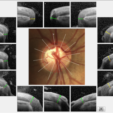
10 Layer Visualization
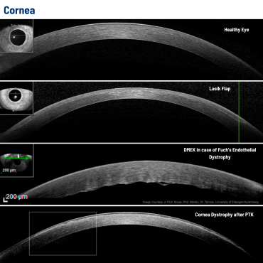
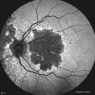
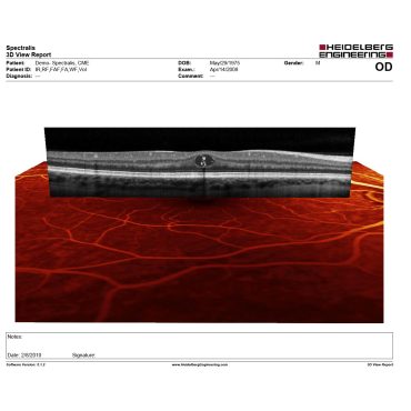
SPECTRALIS® provides you with high resolution OCT images for visualization of 10 retinal layers, to help you confidently describe and pinpoint pathology.
Related Products