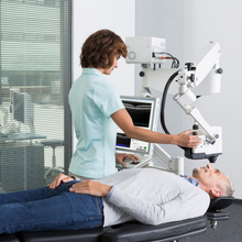
Spectralis OCT PLUS: Similar to the SPECTRALIS OCT, with the exception that it comes with the advanced angiography imaging chassis which allows tilting and panning of the imaging head. The SPECTRALIS OCT PLUS also allows upgrading to angiography imaging.
The SPECTRALIS OCT PLUS features Heidelberg’s unique TruTrack™ active eye tracking and simultaneous dual-beam imaging technologies in an economical, easy to use package. These features allow imaging with enhanced anatomical details, 1 micron measurable change and automatic rescan at follow-up which automatically places follow-up scans in precisely the same location, bypassing operator variability.
The model combines spectral-domain OCT with confocal scanning laser fundus imaging. Additional modules built upon this base platform allow clinicians to tailor a system to the current needs of the practice. As the practice grows, upgrade options allow the SPECTRALIS system to grow along with it. BluePeak™ blue laser autofluorescence, MultiColour™, Anterior Segment, WideField, UltraWideField, and angiography (FA and ICGA) imaging modalities can be added to the SPECTRALIS OCT PLUS when required.
SPECTRALIS OCT also includes Heidelberg’s RNFL and Posterior Pole Asymmetry Analysis for glaucoma diagnosis. These analysis reports are enhanced by the exceptional repeatability of the SPECTRALIS OCT so changes as small as 1 micron can be identified and glaucoma can be confidently diagnosed and managed.
FEATURES:
- TruTrack™ Active Eye Tracking: simultaneously images the eye with two beams of light. One beam captures an image of the retina and maps over 1,000 points to track eye movement. Using the mapped image as a reference, the second beam is directed to the desired location despite blinks or saccadic eye movements.
- Heidelberg Noise Reduction™: combines multiple images captured in the same location, filters out random speckle noise, and retains only data common to the entire set of images. This process retains data reflected from physical structures while mitigating image noise, producing higher quality images with finer detail.
- Confocal Scanning Laser Ophthalmoscopy (cSLO): uses laser light instead of a bright flash of white light to illuminate the retina. The result is a sharp, high contrast image of the object layer located at the focal plane.
- AutoRescan™: automatically places follow-up scans in precisely the same location as the baseline scan.
- FoDi™ fovea-to-disc alignment: a unique alignment technology that automatically tracks and anatomically aligns circle scans to the patients anatomy to improve accuracy and reproducibility of RNFL measurements.
- Multi-modality Imaging Upgrades Available: BluePeak™ blue laser autofluorescense, MultiColour™ Imaging, Anterior Segment, and Widefield imaging modules are available.


