The window to the brain – Gain new insights into neurodegeneration
The Nsite Analytics Module provides a window into the brain with the ability to measure both neuronal and axonal projection within the retina from the central nervous system (CNS). Allowing to monitor changes in the retina of patients with optic neuropathies and neurodegenerative processes including, but not limited to Multiple Sclerosis, Parkinson’s Disease and Alzheimer’s Disease.
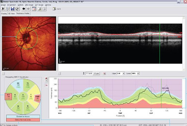
The Nsite Analytics Module provides a comprehensive analysis of the peripapillary retinal nerve fiber layer (RNFL), a unique tissue of the CNS as it contains axons and glia in absence of myelin. Nsite implements a color scheme classification in its reference database to indicate both axonal loss and edematous changes in RNFL. The Nsite Analytics Module also offers macular retinal thickness and posterior pole asymmetry analysis. The macular ganglion cell layer thickness measurements and associated maps indicate whether the observed macular changes are directly related to retinal ganglion cells.
Tracking small changes may be helpful in making better informed clinical decisions. SPECTRALIS® Nsite Analytics Module is able to track small amounts of change to the retinal ganglion cell and axons by minimizing motion artifacts and automatically scanning in the same location during follow-up.
FEATURES:
Clinical Value
- Assessment of neuroaxonal injury and detection of minute changes over time
- High clinical relevance correlating with established clinical criteria
- Associated with physical disability (EDSS) and quality-of-life scores
- Correlated with brain MRI lesions (T1 and T2) and atrophy (BPF)
- Related to disease activity, duration and cognitive decline
- RNFL and ganglion cell thinning can be detected in MS patients with and without optic neuritis
- Excellent reproducibility and sensitivity
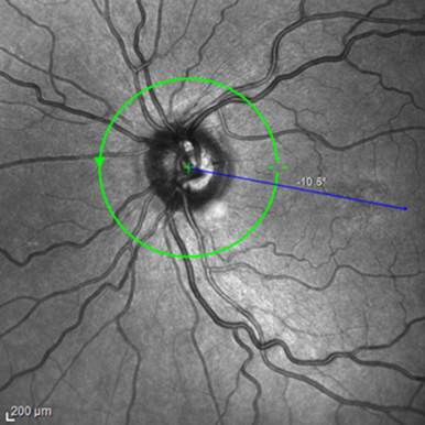
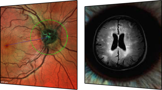
FoBMOC: Automatic Fovea-to-BMO-Center Axis Alignment
Studies have shown that RNFL thinning in MS patients with a history of acute optic neuritis is more likely to be found in the temporal quadrant. The papillomacular bundle (PMB) may be most sensitive to this type of axonal damage. FoBMOC: Automated Fovea-to-BMO-Center Axis Alignment correctly orients the anatomy for PMB measurement accuracy.
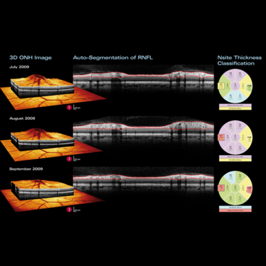
Acute Optic Neuritis
The patient series shows an MS patient with an episode of acute optic neuritis. In the first stage, the optic nerve edema shows no apparent effect on the retinal nerve fiber layer thickness despite the edema. As the edema begins to subside at the second examination, thinning of RNFL in the temporal sector is revealed. By the third visit, only two months later, both the temporal sector and the papillomacular bundle reveal the permanent damage to the axons.
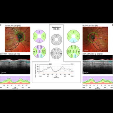
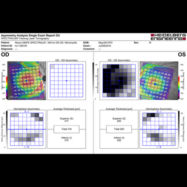
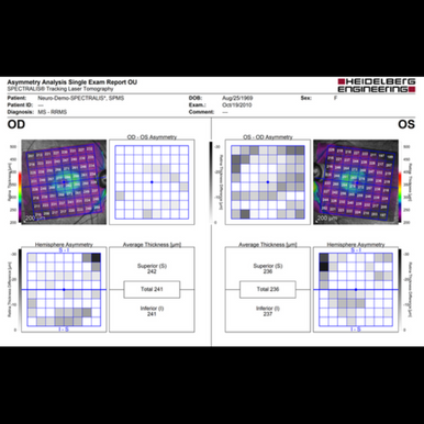
Posterior Pole Asymmetry Analysis
Macular thinning is associated with visual, physical Expanded Disability Status Score (EDSS) as well as other clinical disabilities and may provide insight into neuronal loss and neurodegeneration in MS patients. The Nsite Analytics Module offers a posterior pole asymmetry analysis that precisely measures and maps retinal thickness across the entire posterior pole. A unique asymmetry analysis enables comparison of superior and inferior hemispheres of the same eye and between both eyes. Combined with Nsite retinal nerve fiber layer (RNFL) measurements and the PMB analysis, the posterior pole asymmetry analysis offers a wholistic picture of retinal neurodegeneration and change.
Axonal Follow-up
Tracking small changes may be helpful in making better informed clinical decisions. SPECTRALIS® is able to track minutes changes to the retinal ganglion cell axons by minimizing motion artifacts and automatically scanning in the same location during follow-up. Narrowing the range of variability using TruTrack Active Eye Tracking is key to providing a measurable change of only 1 micron.
Related Products