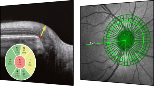
Next generation glaucoma diagnostics

The SPECTRALIS® Glaucoma Module Premium Edition combines the proprietary Anatomic Positioning System (APS) with a series of unique scan patterns to assess the optic nerve head, the retinal nerve fiber layer, and the macular ganglion cell layer. These APS-based scan patterns are automatically matched to each eye’s unique anatomic landmarks and to the characteristics of fine anatomic structures relevant in glaucoma diagnostics.
The Glaucoma Module Premium Edition compares patients’ eyes to a reference database of normal eyes, noting even very small deviations. The precision of the SPECTRALIS AutoRescan function allows confident identification and monitoring of structural changes from visit to visit.
FEATURES:
- Anatomic Positioning System: The patented Anatomic Positioning System (APS) creates an anatomic map of each patient’s eye using two fixed, structural landmarks: the center of the fovea and the center of Bruch’s membrane opening. With APS, all scan protocols are automatically oriented according to the patient’s anatomic map. This enables precise examination of relevant structures and ensures accurate comparisons with reference data, allowing for a highly sensitive assessment of structural change.
- Optic Nerve Head
- Retinal Nerve Fiber Layer
- Posterior Pole


