OPHTHALMIC

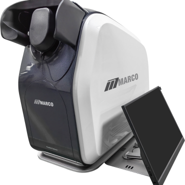
Reticam 3100 Plus Ultra-Widefield Fundus Camera

Brand: Marco

SKU: FCMARETICAM3100

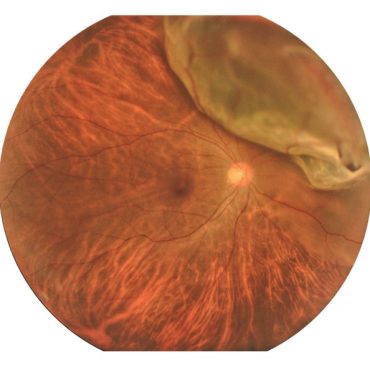
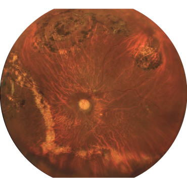
Discover the RetiCam 3100 Plus. See the Periphery. Trust the Colour. A broader retinal view can reveal a more complete clinical picture. RetiCam delivers single-shot ultra-widefield imaging with colour you can trust and extended coverage up to 220°.* *220° field of view achieved through montage stitching and may vary by clinical and operational factors.

Read full product description

A broader view of the retina

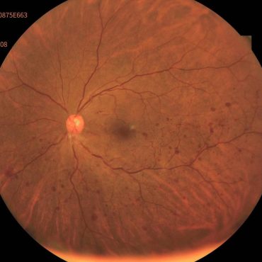
Standard fundus photography primarily visualizes the posterior pole. The RetiCam 3100 Plus extends retinal visualization from the macula through to the far periphery, providing additional clinical context beyond a conventional field of view.

RetiCam provides single-shot ultra-widefield imaging up to 176°. Its montage system extends retinal coverage to up to a 220° field of view,* supporting broader visualization of the retinal periphery.

True-colour clinical visualization

Advanced LED white-light illumination with a continuous spectrum produces true-colour, high-definition retinal images for accurate clinical visualization.

Selectable colour-channel views provide additional ways to examine retinal structures:

  • Choroidal red filter: Highlights deeper retinal and choroidal structures
  • Red-free green filter: Highlights retinal vessels and intraretinal features
  • Superficial blue filter: Highlights superficial retinal structures

Automated image capture

Automated operation helps simplify image acquisition and support an efficient clinical workflow.

Automated functions include:

  • Auto-align
  • Auto-focus
  • Auto-exposure
  • Auto-capture

The system captures images in less than 60 milliseconds and supports up to 16 frames per second.

Non-mydriatic ultra-widefield imaging

The RetiCam 3100 Plus provides non-mydriatic ultra-widefield imaging with a minimum pupil diameter of 3 mm in small-pupil mode.

Soft-exposure technology minimizes pupil constriction, supporting consistent consecutive captures and a comfortable imaging experience. Non-mydriatic imaging complements applicable dilated examinations, established clinical guidelines and professional judgment.

Flexible viewing and connectivity

A web-based viewer provides multi-station access with no per-licence fees, allowing authorized users to review images across compatible workstations.

Additional software and connectivity capabilities include:

  • Gamma, contrast, brightness and colour adjustment
  • Lesion-area and cup-to-disc ratio measurement
  • DICOM 3.0
  • Direct PACS connection
  • Real-time LAN data sharing
  • USB and FTP connectivity
  • JPG, TIFF and DICOM output

Key features

  • Up to a 220° field of view*
  • Single-shot ultra-widefield imaging up to 176°
  • True-colour high-definition imaging for accurate clinical visualization
  • Non-mydriatic ultra-widefield imaging
  • Auto-align, auto-focus, auto-exposure and auto-capture
  • Web-based viewer with multi-station access
  • No per-licence viewer fees
  • Selectable red, green and blue colour-channel views
  • Soft-exposure technology
  • 3 mm minimum pupil diameter in small-pupil mode
  • Compact footprint for pretest rooms and space-constrained clinics
  • DICOM 3.0 and direct PACS connectivity
  • Five-year service plan

Product specifications

Feature Specification
Field of view Single-shot ultra-widefield imaging up to 176°; up to a 220° field of view*
Imaging type Non-mydriatic, true-colour ultra-widefield
Illumination Advanced continuous-spectrum LED white light
Minimum pupil diameter 3 mm in small-pupil mode
Capture speed Up to 16 frames per second; less than 60 ms per capture
Automatic functions Auto-align, auto-focus, auto-exposure and auto-capture
Software tools Gamma, contrast, brightness and colour adjustment; lesion-area and cup-to-disc ratio measurement
Viewer Web-based viewer with multi-station access and no per-licence fees
Connectivity DICOM 3.0, LAN, USB and FTP
Integration Direct PACS connection and real-time LAN data sharing
Output JPG, TIFF and DICOM
Dimensions 19.69 × 9.84 × 18.50 inches (H × W × D)
Service coverage Five-year service plan

Ultra-widefield imaging within reach

The RetiCam 3100 Plus delivers the clarity, efficiency and consistency that modern practices demand. Its combination of peripheral retinal coverage, true-colour imaging, automated operation and accessible positioning makes ultra-widefield imaging easier to introduce into a wider range of practices.

Contact INNOVA to learn more, request a quote or schedule a demonstration.

*220° field of view achieved through montage stitching and may vary by clinical and operational factors.

For qualified medical professional use only. Refer to the User Manual for Canadian authorized indication for use, cautions and precautions.