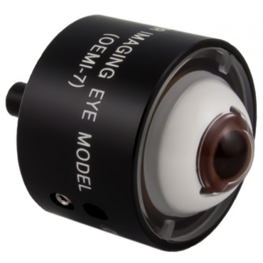
Designed to accurately simulate human eye. Model includes natural surfaces of human eye including anterior chamber and crystalline lens Every effort has been made to duplicate pathological problems found in the human eye. Provides a stable fixed model for evaluation and training. Arteries emanate from the disc with a fluorescent character allowing simulated fluorescein imaging Optic disc has some fluorescent qualities Designed for use with ocular fundus imaging systems such as slit lamps, binocular indirect ophthamoscopes (BIO), fundus cameras and scanning laser ophthalmoscopes (SLO). A peg on the back fits into the Ocular Eye Model Bracket (OEMB1 or OEMB3) which can be attached to any slit lamp. The eye has a retinal detachment showing an elevated retina and retinal tear. It also displays a foreign body, optic disc and blood vessels. A line at the 180 degree meridian designates the region of the equator.
Related Products