TECHNOLOGIES

Optical Coherence Tomography RS-1 Glauvas

Brand: NIDEK

SKU: HA1NKRS-1OCTAz

Optical Coherence Tomography RS-1 Glauvas: Faster workflow without compromising diagnostic confidence

Read full product description

Faster workflow without compromising diagnostic confidence

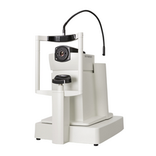
The OCT RS-1 GLauvas from NIDEK is a sophisticated optical coherence tomography device designed to enhance the precision and efficiency of glaucoma diagnosis and management. Utilizing advanced imaging technology, the RS-1 GLauvas delivers high-resolution cross-sectional and three-dimensional images of the retina, allowing clinicians to detect and monitor subtle changes in retinal structure with exceptional accuracy. The device’s intuitive interface and user-friendly features streamline the diagnostic process, making it an essential tool for eye care professionals dedicated to delivering superior patient care. Whether employed in routine screenings or complex case evaluations, the OCT RS-1 Glauvas is a reliable and innovative solution in the field of ophthalmology.

FEATURES

  • 250,000 A-scans/s high-speed imaging:

The incorporation of 250,000 A-scans/s accelerates your workflow by reducing capture time. The high-speed imaging also addresses patient fixation errors thus contributing to greater image clarity and patient comfort.

  • Wide, deep, high-resolution imaging:

With RS-1 Glauvas, a single B-scan image clearly presents the area from the optic nerve head to the temporal vascular arcade, and the 4.2 mm depth B-scan imaging readily captures the oblate retinal shape of myopic eyes. Improvements in AngioScan OCT-Angiography include wider and clearer images for assessing chorioretinal microvasculature.

  • Glaucoma analysis in myopia:

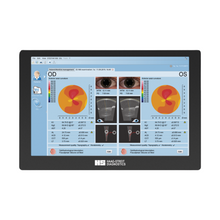
The long axial length normative database*1 presents analysis with axial length compensation, allowing for a more accurate glaucoma assessment in patients with axial myopia. The OCT Viewer automatically switches to this database as required, by using the axial length*2 which is a parameter for scan width correction.

  • Less false positives with deep learning segmentation (DL segmentation)

The accuracy of segmentation affects the outcomes of glaucoma analysis. DL segmentation reduces artifacts and errors in the normative database and thickness maps even in eyes with opacities, thus decreasing false positives and enhancing clinic efficiency by reducing unnecessary follow-up visits. Additionally, the scan width correction allows precise analytics based on the patient’s axial length*2.

  • Structural Normality Map (SN Map) improving diagnostic confidence on early detection

The DL segmentation provides an SN Map that presents structural abnormalities and changes. This functionality aids clinicians in detecting minute structural changes at a glance, contributing to greater diagnostic confidence even for early signs of retinal changes.