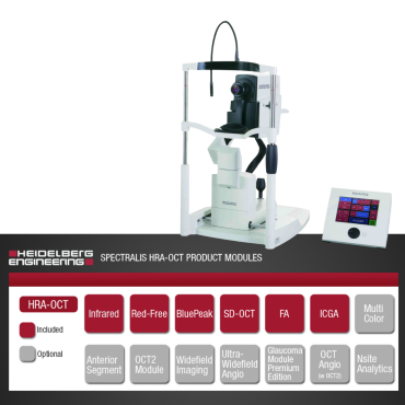
Spectralis HRA-OCT: The current iteration of Heidelberg’s original HRA angiography platform with the addition of SD-OCT. Designed for angiographies, the SPECTRALIS HRA-OCT is built on a Confocal Scanning Laser Ophthalmoscopy (cSLO) platform that is exceptionally sensitive to the fluorescence of Fluoresceine or ICG dyes. This provides focused and high contrast imaging either as still images or Heidelberg’s unique high speed imaging (movies).
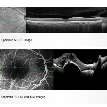
The SPECTRALIS HRA-OCT features SD-OCT imaging with Heidelberg’s unique TruTrack™ active eye tracking and simultaneous dual-beam imaging technologies to allow imaging with enhanced anatomical details, 1 micron measurable change and automatic rescan at follow-up which automatically places follow-up scans in precisely the same location, bypassing operator variability.
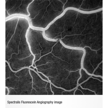
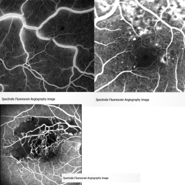
The confocal scanning laser ophthalmoscope (cSLO) technology enhances fluorescein angiography (FA) in two key ways. First, a precise laser wavelength generates the peak emission of light from fluorescein dye, increasing the signal-to-noise ratio. Secondly, confocal technology blocks out-of-focus and scattered light. The results are dramatic. SPECTRALIS® video FA provides clear, detailed images of the capillary bed in real-time and can reveal leakage that may be missed with static FA.
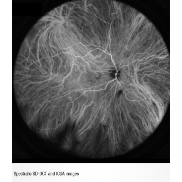
Indocyanine green angiography (ICGA) enables imaging of the choroidal circulation below the retinal pigment epithelium (RPE). ICGA complements fluorescein angiography (FA), which captures blood flow above the RPE. Although ICG angiography is not a replacement for fluorescein, it provides adjunctive information which assists in defining the choroidal circulatory involvement in retinal pathology. Like with FA, SPECTRALIS HRA generates peak emission of light from the ICG dye and the cSLO camera captures clear detailed images of the choroidal vasculature.
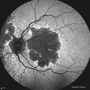
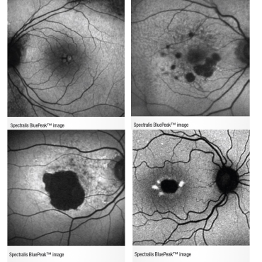
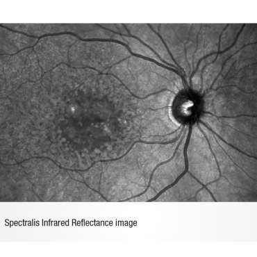
BluePeak™ Blue Laser Autofluorescence imaging takes advantage of the natural fluorescent properties of lipofuscin. BluePeak™ autofluorescence captures fundus autofluorescence (FAF) images, providing both structural and metabolic information about the retina.
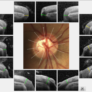
SPECTRALIS HRA-OCT also includes Heidelberg’s RNFL and Posterior Pole Asymmetry Analysis for glaucoma diagnosis. These analysis reports are enhanced by the exceptional repeatability of the SPECTRALIS HRA-OCT so changes as small as 1 micron can be identified and glaucoma can be confidently diagnosed and managed.
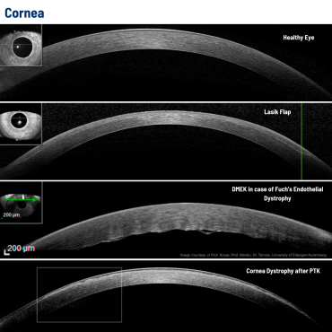
Like all SPECTRALIS models, the SPECTRALIS HRA features an upgradable, modular design: allowing configuration of each SPECTRALIS to the specific diagnostic workflow in the practice or clinic. Upgrade options include: MultiColor™, widefield, ultra-widefield and anterior segment imaging.
FEATURES:
- TruTrack™ Active Eye Tracking: simultaneously images the eye with two beams of light. One beam captures an image of the retina and maps over 1,000 points to track eye movement. Using the mapped image as a reference, the second beam is directed to the desired location despite blinks or saccadic eye movements.
- Heidelberg Noise Reduction™: combines multiple images captured in the same location, filters out random speckle noise, and retains only data common to the entire set of images. This process retains data reflected from physical structures while mitigating image noise, producing higher quality images with finer detail.
- Confocal Scanning Laser Ophthalmoscopy (cSLO): uses laser light instead of a bright flash of white light to illuminate the retina. The result is a sharp, high contrast image of the object layer located at the focal plane.
- High Resolution Angiography Imaging: Exceptional detail is obtained by the SPECTRALIS HRA’s cSLO imaging device.
- High Speed Angiographies: SPECTRALIS HRA’s cSLO technology is not limited like the flash imaging technology so multiple images can be acquired within a second. This allows movies to be acquired where each frame is a full resolution image.
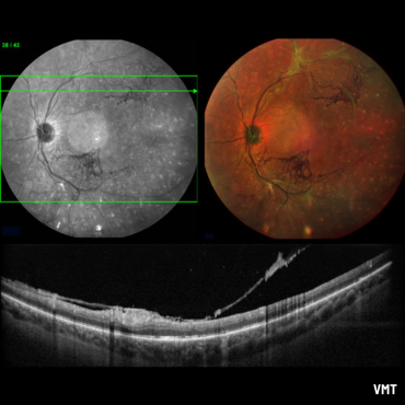
- Dual Imaging Capabilities: SPECTRALIS HRA’s dual imaging capabilities allows side-by-side FA and ICGA angiographies. This unique capability provides valuable insights provided by these different imaging modes.
- Autocomposite Imaging: The SPECTRALIS HRA’s panning head and live composite imaging allows expansion of the field easily for any technician.
- AutoRescan™: automatically places follow-up scans in precisely the same location as the baseline scan.
- FoDi™ fovea-to-disc alignment: a unique alignment technology that automatically tracks and anatomically aligns circle scans to the patients anatomy to improve accuracy and reproducibility of RNFL measurements.
- Multi-modality Imaging Upgrades Available: MultiColour™ Imaging, Anterior Segment, Widefield and Ultra-Widefield imaging modules are available.
Related Products